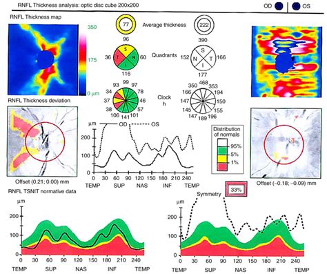
optical coherence tomography thickness measurements|optical coherence tomography test results : fabrication We sought to compare the retinal thickness measurements collected using different optical coherence tomography (OCT) devices. This prospective study included 21 .
webGaranta a sua segurança, mantenha a revisão da sua moto sempre em dia! Entre em contato com Eltinho Motos e Yamasul e traga sua moto para um orçamento. Venha nos visitar: Rua Professora Luiza Maraninche 809 no Bairro: Centro Cidade de Camaquã, RS. O Cep de nosso endereço é: 96180-000.
{plog:ftitle_list}
WEB2 de fev. de 2023 · #paqueta #dance #songCopyright Disclaimer under section 107 of the Copyright Act of 1976, allowance is made for “fair use” for purposes such as criticism, co.
The CPT is based on the intersection of six radial line scans (compared with 128 macular thickness measurements made for the CSMT). The CPT has a ± indicating the deviation of the measurement. A deviation greater than 10 percent of the CPT implies an .This allows your ophthalmologist to map and measure their thickness and .Optical coherence tomography has emerged as a useful imaging technique by providing new high-resolution cross-sectional information about various pathological features of the macula. .
moistur meter for fire wood
Optical coherence tomography (OCT) is now commonly used to measure macular thickness quantitatively for disease diagnosis and to monitor the efficacy of therapeutic modalities. 15–24 Optical coherence tomography . Optical coherence tomography (OCT) is a non-contact imaging technique which generates cross-sectional images of tissue with high resolution. Therefore it is especially valuable in organs, where traditional microscopic . We sought to compare the retinal thickness measurements collected using different optical coherence tomography (OCT) devices. This prospective study included 21 .The six OCT systems provided different results for CRT. The measurements with the Stratus OCT showed the lowest thicknesses, whereas those with the Cirrus HD-OCT and Spectralis .
This allows your ophthalmologist to map and measure their thickness and changes over time. These measurements help with diagnosis. They also guide treatment for glaucoma as well as retinal disease, like age .
Optical coherence tomography (OCT) segmentation boundaries of retinal thickness according to different OCT devices. ( a ) Segmentation boundaries of PLEX Elite, . An optical coherence tomography test is a quick and easy imaging test that can help your provider see to the back of your eyeball. It provides three-dimensional images of the . Optical coherence tomography (OCT) is a noninvasive imaging technique that uses visible and infrared electromagnetic waves to provide detailed, cross-sectional images .
Background To compare the repeatability and reproducibility of corneal and corneal epithelial thickness mapping using anterior segment optical coherence tomography (AS-OCT) according to tear film break-up time (TBUT). Methods The included eyes were divided into three subgroups according to TBUT (group 1: TBUT ≤ 5 s, group 2: 5 s < TBUT ≤ 10 s, and group 3: .dierent optical coherence tomography devices Ki Tae Nam1, CheolminYun2*, Myungho Seo1, Somin Ahn2 & Jaeryung Oh2 We sought to compare the retinal thickness measurements collected using dierent . Optical coherence tomography (OCT) is a real-time, in-situ, non-invasive imaging device that is able to perform a cross-sectional evaluation of tissue microstructure based on the specific intensity of back-scattered and reflected light. . OCT measurements of epithelial thickness were performed in 28 healthy patients at six different locations .Traditional methods for evaluating macular edema, such as slitlamp biomicroscopy, stereoscopic photography, and fluorescein angiography, are relatively insensitive to small changes in retinal thickness and are qualitative at best. 4 The introduction of optical coherence tomography (OCT) has enabled clinicians to reliably detect and measure .
Blumenthal EZ, Williams JM, Weinreb RN, Girkin CA, Berry CC, Zangwill LM. Reproducibility of nerve fiber layer thickness measurements by use of optical coherence tomography. Ophthalmology. 2000;107:2278–2282.
We sought to compare the retinal thickness measurements collected using different optical coherence tomography (OCT) devices. This prospective study included 21 healthy cases, and the retinal thickness was measured using the PLEX Elite (Carl Zeiss Meditec, Dublin, California, USA), DRI OCT-1 Atlantis (Topcon Corp, Tokyo, Japan), Cirrus 5000 HD .Purpose: To analyze retinal thickness and volume measurements of X-linked retinoschisis patients by spectral-domain optical coherence tomography (SD OCT) and correlate these findings with visual acuity and patient age. Design: Retrospective comparative case series. Methods: Sixty-three eyes of 33 male patients with X-linked retinoschisis were gleaned from a .Central subfield mean thickness is the preferred OCT measurement for the central macula because of its higher reproducibility and correlation with other measurements of the central macula. Total macular volume may be preferred when the central macula is less important. . To evaluate optical coherence tomography (OCT) measurements and methods .
: A custom-built spectral domain optical coherence tomography system comprising a broadband titanium:sapphire laser operating at 800 nm and a high-speed charge coupled device (CCD) camera with a read-out rate of 47 kHz was used for measurement of precorneal tear film thickness. The system provides a theoretical axial resolution of 1.2 μm in .
Optical coherence tomography (OCT) is now commonly used to measure macular thickness quantitatively for disease diagnosis and to monitor the efficacy of therapeutic modalities. 15–24 Optical coherence tomography measurements of macular thickness have been demonstrated to be highly reliable. 25–29 However, there are limited data of normal .A major reason for this is probably that macular thickness measurement did not take advantage of the improved resolution since the ILM and RPE were the most clearly identifiable layers, even on the lower resolution OCT images. . Hertzmark E, et al. Optical coherence tomography measurement of macular and nerve fiber layer thickness in normal .
Importance: Understanding measurement variability and relationships between measurements obtained on different optical coherence tomography (OCT) machines is critical for clinical trials and clinical settings. Objective: To evaluate the reproducibility of retinal thickness measurements from OCT images obtained by time-domain (TD) (Stratus; Carl Zeiss Meditec) . This study examines the measurement of film thickness, curvature, and defects on the surface or inside of an optical element using a highly accurate and efficient method. This is essential to ensure their quality and performance. Existing methods are unable to simultaneously extract the three types of information: thickness, curvature, and defects. Spectral-domain .Optical coherence tomography (OCT) is a non-invasive imaging technology that uses low-coherence light to obtain cross-sectional images within biological samples. It is widely used in the field of ophthalmology to measure retinal thickness, for instance. The images generated are similar to ultrasound images in appearance.Purpose : The portable Retinal Thickness Module (RTM, Kubota Vision) is a novel home-based patient- administered optical coherence tomography (OCT) device to measure retinal thickness and identify macular edema. This .
Purpose: Compare the use of optic disc and macular optical coherence tomography measurements to predict glaucomatous visual field (VF) worsening. Methods: Machine learning and statistical models were trained on 924 eyes (924 patients) with circumpapillary retinal nerve fiber layer (cp-RNFL) or ganglion cell inner plexiform layer (GC . An intravascular optical coherence tomography (IVOCT) method was developed to simultaneously measure the circumferentially distributed intima-media thickness, strain and strain rate of carotid artery with high accuracy in vivo.. An elastic modulus calculation method was proposed based on the cyclic strains for biomechanical properties characterization of the . In Vivo Intraocular Lens Thickness Measurement and Power Estimation Using Optical Coherence Tomography. Ehsan Barzanouni, MD, 1 , 2 Diba Idani, . Optical Coherence Tomography Measurements. OCT scans were performed using anterior segment module, (Topcon 3D OCT-1000 Topcon Corporation, Tokyo, Japan) after pupillary dilation. . Optical coherence tomography (OCT) is a real-time, in-situ, non-invasive imaging device that is able to perform a cross-sectional evaluation of tissue microstructure based on the specific intensity of back-scattered and reflected light. . OCT measurements of epithelial thickness were performed in 28 healthy patients at six different locations .
moisture activity meter
To compare a time-domain (Stratus) and a spectral-domain (Spectralis) optical coherence tomography (OCT) device in assessing foveal thickness in healthy subjects. In this observational study 40 .Purpose: To evaluate the reproducibility of nerve fiber layer (NFL) thickness measurements by optical coherence tomography (OCT) in individuals with silicone oil-filled eyes. Methods: Eighteen patients who had undergone pars plana vitrectomy and silicone oil tamponade for retinal detachment were enrolled in a prospective, case-controlled clinical study. Although the clinical assessment of enamel thickness is important, hardly any tools exist for accurate measurements. The purpose of this study was to verify the precision of enamel thickness measurements using swept-source optical coherence tomography (SS-OCT). Human extracted maxillary central and lateral incisors were used as specimens. Twenty-eight . Optical coherence tomography (OCT) has been applied to measure peripapillary retinal nerve fiber layer (RNFL) thickness at a micrometer scale in several optic neuropathies such as non-arteritic .
Purpose: To evaluate the reproducibility of central subfield thickness (CST) and volume measurements from optical coherence tomography (OCT) images obtained with Zeiss Stratus and Optovue RTVue, and formulate equations to convert these measurements from RTVue to 'equivalent' Stratus values. Methods: Cross-sectional observational study from 309 . We sought to compare the retinal thickness measurements collected using different optical coherence tomography (OCT) devices. This prospective study included 21 healthy cases, and the retinal thickness was measured using the PLEX Elite (Carl Zeiss Meditec, Dublin, California, USA), DRI OCT-1 Atlantis (Topcon Corp, Tokyo, Japan), Cirrus 5000 HD .
Introduction. The arrival of Optical Coherence Tomography (OCT) has changed the way that retinal pathology is diagnosed and managed. OCT imaging allows non-invasive cross-sectional imaging of the human retina [].Good correlation with retinal histology [2–4] pertains OCT technology to the clinical diagnosis of a variety of ocular pathologies [5–8] based .Purpose: To obtain retinal nerve fibre layer thickness measurements by optical coherence tomography (OCT) in normal Indian population. Materials and methods: Total of 118 randomly selected eyes of 118 normal Indian subjects of both sex and various age groups underwent retinal nerve fiber layer thickness analysis by Stratus OCT 3000 V 4.0.1.
optical coherence tomography test results

moisture air flow meter
optical coherence tomography test cost
optical coherence tomography review
webE-mail. Senha. Esqueceu a senha? Acessar. Criar conta.
optical coherence tomography thickness measurements|optical coherence tomography test results