measurement of small intestine thickness|The Radiology Assistant : US of the GI tract : department Store The 3-6-9 rule is a simple aide-memoire describing the normal bowel caliber: small bowel: <3 cm. large bowel: <6 cm. appendix: <6 mm. cecum: <9 cm. Above these dimensions, the bowel is . Resultado da Hotel Panamby São Paulo. 8.8 - Excelente ( 2080) 1.6 km até Arena Palestra Itália – Allianz Parque. R$262 por diária. Preço previsto para: 24 de ago. - 25 de ago. Comparar preços. Hotel.
{plog:ftitle_list}
Resultado da NINFETINHAS. O melhor grupo do Telegram +18 com conteúdos amadores , vazados do whatsapp ou da internet inteira, videos de famosas, grupos .
Normally, single small bowel wall thickness during compression is about 1.5 - 2.5 mm. Measuring bowel wall thickness by US this way is reproducible and comparable to what surgeons do with their fingers during laparotomy to decide whether small bowel is abnormal.The 3-6-9 rule is a simple aide-memoire describing the normal bowel caliber: small bowel: <3 cm. large bowel: <6 cm. appendix: <6 mm. cecum: <9 cm. Above these dimensions, the bowel is . Bowel wall thickness is the most important feature in intestinal ultrasound assessment and an important parameter used in detecting intestinal disease. Thickness is determined by measuring all of the wall strata from the . Eleven studies met the inclusion criteria and reported small intestinal length measurement using four imaging modalities: barium follow-through, ultrasound, computed tomography, and magnetic resonance.

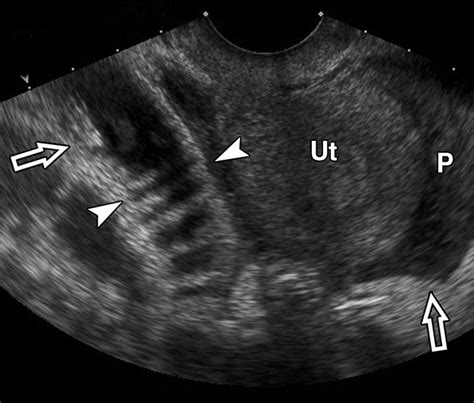
An ultrasound image showing 2 sections of small intestine. Two callipers (crosses with linked dotted line) have been placed to measure intestinal wall thickness, from the inner edge of . Transvaginal ultrasound image shows complex fluid compatible with pus (P) surrounding uterus (Ut). Adjacent small-bowel loop is dilated and thick-walled (arrowheads), reflecting reactive enteritis and ileus.Abstract. Purpose: Small intestinal length has prognostic significance for patients with short bowel syndrome, and accurate measurement of Roux-en-Y limbs is considered important. .Purpose: To assess the normal small bowel parameters, namely bowel diameter, bowel wall thickness, number of folds (valvulae connivientes) per 2.5 cm (in.), fold thickness and .
Purpose: To describe the gastrointestinal (GI) wall thickness and the thickness of individual wall layers in healthy subjects using ultrasound and to determine whether demographic factors, the . These images are of a patient with a closed loop small bowel obstruction. Notice the group of small bowel loops with a thickened wall in the right upper abdomen (yellow arrow). The mesenteric edema (red arrow) . Abdominal ultrasonography can be a useful tool in the diagnostic work up of small animals affected by gastrointestinal diseases. 1–3 The ultrasonographic appearance of the normal feline gastrointestinal tract has . Although not shown here, there was diffuse selective thickening of the muscularis layer affecting the remainder of the small intestine. The final diagnosis was inflammatory bowel disease . normal intestinal wall .
Ultrasound Imaging of Bowel Pathology: Technique
A thinner small intestinal wall compared to the present findings has been reported scanning ex vivo intestine using a 7.5 MHz probe, 9 and also a high frequency (10–18 MHz) probe. 8 Noteworthy, in the present study, the thicker US measurement was in line with a thicker histological measurement of the wall, compared to the findings previously .
maximum thickness of 4 mm on the transverse view.13 In normal dogs and cats, the small intestines are relatively uniform in distribution. Depending on the segment of small intestine, some layers may be thicker than others. This can be used to identify the different segments of intestines. For example, in the dog, the mucosal layer of theThe duodenal wall thickness was significantly greater than other parts of the small intestine, and this difference was accentuated by sedation (awake mean 2.4 mm; sedated mean 2.71 mm). The mean small intestinal wall thickness was 2.1 .
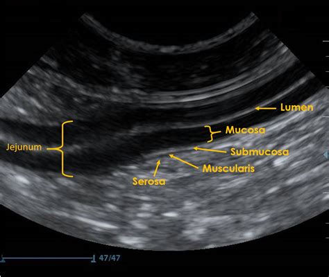
The normal sonographic thickness of the individual layers (ie, mucosa, submucosa, muscularis and subserosa-serosa) of the intestinal wall was evaluated in 20 clinically healthy cats. The mean thickness of the wall was 2.20, 2.22, 3.00 and 2.04 mm for duodenum, jejunum, ileum (fold) and ileum (betwee . 2.2.1. Intestinal wall thickness measurements . For the same transverse section of small intestine from a single cat, each observer was instructed to measure the thicknesses of the intestinal wall layers and segment the area (Figure 1). For US images that included more than one transverse section, a reference region of interest was created .The use of ultrasound to measure small bowel thickness is an important part of any ultrasound examination of the abdomen. Increased thickness of the intestinal wall is a hallmark for the detection of diseases ranging from inflammatory bowel disease to neoplasia. Our subjective impression has been th .The results would provide the information necessary to decide whether measurement of ultrasonographic wall thickness can predict IBD in dogs. Methods: The intestinal wall thickness of 75 dogs with idiopathic IBD, as measured by ultrasonography, was compared with recently published normal values. IBD was either confirmed histologically (n = 54 .
Ultrasonography of the gastrointestinal tract
Noninvasive measurement of small intestine length is challenging. Three-dimensional imaging modalities reduce the risk of length underestimation, which is common with two-dimensional techniques. . T1-weighted signal, Spin echo time: 9.8 ms, field-of-view: 25 x 25, slice thickness: 3 mm; mice anesthetized-Ex vivo measure: 0 d: 2: Dillman 2016 35-
Ultrasound can be very useful for assessing the small intestine. Ultrasound examination can provide information regarding bowel thickness, layering pattern, motility and evidence of diffuse or segmental dilation. . Normal wall thickness is generally as follows: Wall thickness: Cats: Dogs: Duodenum <2.8 mm: 3-6 mm: Jejunum <2.5 mm: 2-5 mm .3-6-9 rule (bowel) | Radiology Reference Article | Radiopaedia.org
measurement of fluid film thickness
Intestinal Wall Thickness and Layering The normal wall thickness of the cecum, colon and most of the small intestine is less than 3 mm. The ileum has a more prominent muscular layer and can measure up to 5 mm. Diseases such as Inflammatory bowel disease (IBD) and alimentary lymphoma can cause thickening of the intestine. The use of ultrasound to measure small bowel thickness is an important part of any ultrasound examination of the abdomen. Increased thickness of the intestinal wall is a hallmark for the detection .
Mucosal folds: The inside surface of the small intestine is not flat, but rather is made up of circular folds that increase the surface area.; Intestinal villi: The mucous folds in the small intestine are lined with multitudes of tiny .and/or thickness of intestinal wall involving selectively some intestinal layers have been described in feline gastro-intestinal disorders.6–9 In particular, an ultrasonographical pattern characterised by a diffusely thickened muscularis propria of the small intestine has been associated both with gastrointestinal lymphoma and inflammatory bowelRESULTS: A positive association between intestinal wall thickness in dogs and either the histological diagnosis or the response to treatment was not found. Ultrasonographic intestinal wall measurements do not appear to be able to establish a diagnosis of intestinal inflammation and may result in a false negative diagnosis in cases of IBD.
2.2.1 Intestinal wall thickness measurements. For the same transverse section of small intestine from a single cat, each observer was instructed to measure the thicknesses of the intestinal wall layers and segment the area (Figure 1). For US images that included more than one transverse section, a reference region of interest was created using . Intestinal wall layer thickness measurements of the mucosa, submucosa, muscularis, serosa layer, and total thickness of these layers were performed on five cats with small cell epitheliotropic . Abdominal ultrasonography can be a useful tool in the diagnostic work up of small animals affected by gastrointestinal diseases. 1–3 The ultrasonographic appearance of the normal feline gastrointestinal tract has been described and reference ranges for wall thickness of different segments have been reported. 4,5 Changes in echogenicity and/or thickness of .
The Radiology Assistant : US of the GI tract
Total mucus thickness over time in the rat small intestine before and after removal of the loosely adherent mucus layer. , Duodenum (n = 7); , jejunum (n = 6); , ileum (n = 5 before mucus removal and 8 after mucus removal). Values are means ± SE and represent the total mucus thickness at each time point.The normal sonographic appearance of stomach, small bowel, and colon was determined in normal dogs of small, medium, and large breeds. In all dogs studied, the stomach wall ranged from 3 mm to 5 mm in thickness, and the small and .
The relationship between histological and ultrasonographic thickness of the intestinal wall and its layers in cats is unknown so far. The aims of this study were to establish the relationship between ultrasonographic measurements in the transverse and longitudinal planes of the small intestine and to establish the agreement between ultrasonographic and .Furthermore, Lodwick et al., studied a group of 13 subjects planned to undergo a lengthening procedure for short bowel syndrome (intraoperative small intestine length median: 69 cm, range: 21–170 cm) and found a strong correlation between the intraoperative and radiographic measurements of small intestine length (median 71 cm, range: 29–142 . Here we describe a third approach, ex vivo measurements of mucus adhesion, properties, thickness, and growth, as a function of time in mouse and human colon and mouse small intestine. The principle of measuring mucus thickness is adapted from the in vivo technique where charcoal particles were added onto the mucosal surface and visualized by . Direct maximum bowel wall thickness measurements were also classified as mild (3 to 5 mm), moderate (5 to 9 mm), or severe (>9 mm) based on recent guidance from the American Gastrointestinal Association . The automated measurements of small intestinal structural features relied on bowel segmentation. Both study radiologists independently .
The Radiology Assistant : CT
The Challenge of Small Intestine Length
The results of the normal small intestinal wall thickness derived from one of these two studies (Penninck and others 1989) have since been used as a standard for the identification of increases in wall thickness (Manczur and Vörös 1999, Kull and others 2001, Delaney and others 2003). . The results provided the information necessary to .
Sonography Intestinal Assessment, Protocols, and

Resultado da 10 de nov. de 2023 · O Palmeiras recebe o Internacional, nesta quarta-feira, pela 34ª rodada do Campeonato Brasileiro.O duelo acontece às 21 horas (de Brasília), na Arena Barueri. A transmissão do jogo será feira pelos canais Premiere (pay-per-view) e SporTV. Como chegam os times? O Palmeiras .
measurement of small intestine thickness|The Radiology Assistant : US of the GI tract