analyzing protein content with uv-visible spectroscopy protocol|uv concentration of protein : mail order NPTEL WEB5 de fev. de 2024 · 战雷涂装中文网是国内最大的《warthunder》(战争雷霆)玩家创作研究、交流学习和分享涂装语音包等自定义内容的专业中文社交平台!这里你可以找到最新最好看的涂装,与更多的爱好者交流,与大家一起分享游戏、涂装的乐趣与热爱。
{plog:ftitle_list}
WEBSkill Games: 选择一个免费Skill Games, 尽享快乐时光. Super Fowlst 2 Swingo Crossy Road Space Thing Noob Archer Moto X3M Winter Odd Bot Out Fish Eat Fish Bouncy Dunks Temple Run 2: Holi Festival Super Tunnel Rush Stickjet Challenge Impossible Tic-Tac-Toe Stupid Zombies Poor Bunny Temple Run 2: Jungle Fall Stickman Fight: .
Protein Quantitation using a UV-visible Spectrophotometer. Introduction. Generally, protein quantitation can be made using a simple UV-Visible spectrophotometer. The V-630 Bio (Figure .Protein Quantitation using a UV-visible Spectrophotometer Application Note UV-0003 Introduction Generally, protein quantitation can be made using a simple UV-Visible spectrophotometer. The V-630 Bio (Figure 1) is a UV-Visible spectrophotometer designed for biochemical analysis. The V-630 Bio includes 6 quantitative methods based on UVNPTELUV-Visible Analysis of Bitterness and Total Carbohydrates in Beer Application Note 52467 Key Words Evolution 200, Beer, Bitterness, Brewing, Polyphenols, Quality Control, UV-Visible Spectroscopy Background UV-Visible spectroscopy is one of the most basic techniques for testing a variety of materials. It is routinely used for . protein content .
uv concentration of protein
uv absorption of protein
UV-Vis spectroscopy is used to quantify the amount of DNA or protein in a sample, for water analysis, and as a detector for many types of chromatography. Kinetics of chemical reactions are also measured with UV-Vis spectroscopy by taking repeated UV-Vis measurements over time. UV-Vis measurements are generally taken with a spectrophotometer.In the present study we have reported the protocol from quantitative estimation of BSA content. Firstly using UV-Visible spectroscopy, the excitation wavelength for BSA protein has been identified. Analysis of different functional groups has been carried out using FTIR spectroscopy.The actual value of UV absorbance for a given protein must be determined by some absolute method, e.g., calculated from the amino acid composition, which can be determined by amino acid analysis (4). The UV absorbance for a protein is then .
Generally, protein quantitation can be made using a simple UV-Visible spectrophotometer. The V-730 Bio (Figure 1) is a UV-Visible spectrophotometer designed for biochemical analysis. The V-730 Bio includes 6 quantitative methods based on UV absorption spectrophotometry including the Lowry, Biuret, BCA, Bradford, and WST methods. Figure 1. .
UV-Visible spectroscopy is one of the important ways for the quantitative analysis of protein in any plant sources along with the analysis of the composition of amino acids [16, 17]. This method .Methods using UV-visible spectroscopy. A number of methods have been devised to measure protein concentration, which are based on UV-visible spectroscopy. These methods use either the natural ability of proteins to absorb (or scatter) light in the UV-visible region of the electromagnetic spectrum, or they chemically or physically modify . Types of Detectors in UV-Visible Spectroscopy. In the domain of UV-Visible spectroscopy, detectors play an indispensable role. Their primary function is to convert light into proportional electrical signals, which subsequently determine the spectrophotometer’s response. Delving into the technicalities, there are four primary types of .
what is a rockwell hardness test
scattering protein by uv

what is brinell hardness test
%PDF-1.6 %âãÏÓ 600 0 obj > endobj 624 0 obj >/Filter/FlateDecode/ID[0D68C31A8E0937438E797621B9C54FBD>81B828330A4A3B4A91845ACEAE23EA1A>]/Index[600 45]/Info 599 0 R .into its single-stranded components. As UV-Visible absorption spectroscopy is a non-destructive technique, this analysis can be carried out using samples which can be further analyzed later. In an ideal scenario, the resulting sigmoidal curve, as shown in Figure 3, should be observed, where no change is observed In addition, it can be used to study protein interactions. This protocol details the basic steps of obtaining and interpreting CD data, and methods for analyzing spectra to estimate the secondary .%PDF-1.7 %âãÏÓ 136 0 obj > endobj xref 136 41 0000000016 00000 n 0000001621 00000 n 0000001771 00000 n 0000001815 00000 n 0000003000 00000 n 0000003544 00000 n 0000004164 00000 n 0000004789 00000 n 0000004826 00000 n 0000004874 00000 n 0000005470 00000 n 0000005584 00000 n 0000005673 00000 n 0000005770 00000 n .
Generally, protein quantitation can be made using a simple UV-Visible spectrophotometer. The V-730 Bio (Figure 1) is a UV-Visible spectrophotometer designed for biochemical analysis. The V-730 Bio .
Circular dichroism (CD) spectroscopy has emerged as a powerful tool in the study of protein folding, structure, and function. This review explores the versatile applications of CD spectroscopy in unraveling the . Generally, protein quantitation can be made using a simple UV-Visible spectrophotometer. The V-730 Bio (Figure 1) is a UV-Visible spectrophotometer designed for biochemical analysis. The V-730 Bio . 5. UV SPECTROSCOPY • UV spectroscopy is concerned with the study of absorption of UV radiation which ranges from 200nm to 400nm, colored compounds absorb the radiation from 400nm to 800nm(visible region). • Colorless compounds absorb the radiation at UV region. In both UV spectroscopy and visible spectroscopy, the valence electrons absorb .
Aim: Simple, precise, accurate, cost effective, rapid, and sensitive UV/visible spectrophotometric method was developed at the wavelength of maximum absorption, λmax (290 nm). The developed UV/visible spectrophotometric method was validated as per ICH guidelines for the estimation of Linagliptin in active pharmaceutical dosage form and bulk form.PROTEIN SOLUTIONS CONTAMINATED WITH NUCLEIC ACIDS For a protein solution that is known to be contaminated with nucleic acids the following method can be used to give a reasonable approximation of the protein concentration. This works for nucleic acid content up to 20% w/v or A 280 / A 260 < 0.6 • Zero spectrophotometer with a buffer blank.UV-Visible spectroscopy employs the UV (400200 nm) and visible (800- -400 nm) region of the electromagnetic spectrum. This energy associated with this region is quite high and . • Analysis of Hydrocarbons: In molecules containing long saturated hydrocarbon chains, the corresponding molecular ion peak is observed along with peaks
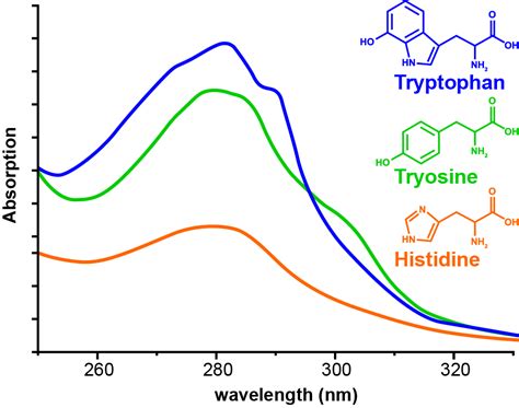
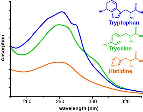
UV–Vis spectroscopy has many uses including detection of eluting components in high performance liquid chromatography (HPLC), determination of the oxidation state of a metal center of a cofactor (such as a heme), determination of the maximum absorbance of proteins for measurement of their concentrations or monitoring of structural changes in .
UV-Vis spectroscopy is a method that can monitor and measure the interactions of UV and visible light with different chemical compounds in the wavelength range between 200 and 780 nm.The technique exploits different physical responses of light and analytes within the sample such as absorption, scattering, diffraction, refraction, and reflection [1].
Raman spectroscopy has a high molecular specificity, making it an excellent technique for materials analysis. However, Raman scattering is a rare phenomenon with an exceptionally low probability .
With traditional methodologies that rely on standard fixed-pathlength UV-visible spectroscopy, the turn-around time may be on the order of hours. . The Solo VPE system has been used to analyze protein solutions at concentrations ranging as high as 300 mg/mL . Its search algorithm looks for an initial response of 1 AU (independent of sample .In the present study we have reported the protocol from quantitative estimation of BSA content. Firstly using UV-Visible spectroscopy, the excitation wavelength for BSA protein has been identified. Analysis of different functional groups has been carried out using FTIR spectroscopy. The electromagnetic spectrum refers to the known frequencies and their linked wavelengths of the known photons [5, 8].Although all the wavelengths shown in Fig. 8.1 can be used for analysis, the range of wavelengths usually employed in spectroscopy is relatively narrow and includes mainly the UV, visible, infrared (IR), ultrasound, and FM radio (nuclear .
what is brinell hardness test used for

This offer is only available to new and eligible customers. Full Offer Terms and Conditions. Qualifying. Promotion runs from 09:00 GMT on 12th January until 23:59 GMT on 14th .
analyzing protein content with uv-visible spectroscopy protocol|uv concentration of protein