measuring melanoma thickness|clark level for melanoma staging : exporter The Breslow thickness describes how thick the melanoma is. It measures in millimetres (mm) how far the melanoma cells have grown down into the layers of skin. There are 5 levels of .
Resultado da FreeBitco.in is a bitcoin dice game with a high cost-to-reward ratio. Add to that the sheer simplicity, speed, and quality of the gameplay and you’re in for a crypto-treat! We have the most popular bitcoin dice game on the internet with more than 37,361,449 registered users, a whopping 108,456,862,013 .
{plog:ftitle_list}
30 de ago. de 2019 · O VStatus é um app gratuito para celulares Android que permite baixar vídeos para colocar nos Status do WhatsApp. O conteúdo inclui vídeos românticos, engraçados, de amor, músicas e .
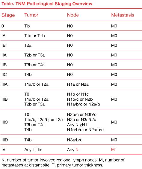
tnm melanoma staging chart
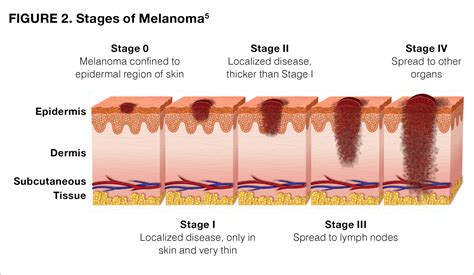
The Breslow Depth is a helpful measure of how far melanoma has invaded the body and the Clark Level is a staging system that describes the depth of melanoma as it grows in the skin.Stage 4 Melanoma. Stage IV melanoma has metastasized (spread) to other .
melanoma stages explained
Melanoma treatments have improved significantly with the addition of .
Knowing the stage of melanoma helps to determine your prognosis and identify .
Tumor thickness: The thickness of the melanoma is called the Breslow measurement. In general, melanomas less than 1 millimeter (mm) thick (about 1/25 of an inch) have a very small chance .
The Breslow thickness describes how thick the melanoma is. It measures in millimetres (mm) how far the melanoma cells have grown down into the layers of skin. There are 5 levels of .
The thickness of a tumor is the most important risk factor. The thicker the tumor, the more likely it will spread. Breslow depth is the tumor’s thickness. It measures how deep below the surface of the skin the melanoma cells have reached. . Measured with a calibrated ocular micrometer. Measurement is rounded to the nearest 0.1 mm. Report as "at least _ mm" if the maximum tumor thickness cannot be .
Accurate staging of cutaneous malignant melanoma depends on the availability of full-thickness biopsy specimens. Sentinel lymph node biopsy, if accessible, should be .
melanoma stages chart
Breslow depth is a measurement (in millimeters) from the surface of the skin to the deepest component of the melanoma. Tumor thickness: Known as Breslow thickness or Breslow depth, this is a significant factor in predicting how far a .Breslow thickness is the measurement of the depth of the melanoma from the surface of your skin down through to the deepest point of the tumour. It’s measured in millimetres (mm) with a .Knowing the stage of melanoma helps to determine your prognosis and identify the best treatment plan for you. Melanoma staging is based on the American Joint Committee on .Breslow thickness is the single most important prognostic factor for clinically localised primary melanoma.1 Breslow thickness is measured from the top of the granular layer of the epidermis .

Cutaneous melanoma is the fifth most common cancer diagnosis in the United States, and the incidence continues to increase with a projected 110000 new cases in the year 2030 compared to around 65000 new cases in 2011.[1] . Measurement of Breslow tumor thickness to 0.1mm is recommended, round down to those ending in decimal places 1 to 4 .Photoacoustic imaging (PAI) is an emerging biomedical imaging technology, which can potentially be used in the clinic to preoperatively measure melanoma thickness and guide biopsy depth and sample location. We recruited 27 patients with pigmented cutaneous lesions suspicious for melanoma to test the .Correlation between melanoma thickness measured using a linear 14-MHz frequency ultrasound sensor and histologically estimated melanoma thickness according to Breslow was lower if melanoma was thinner (1-2 mm). However, in cases of thicker melanoma (>2 mm), very strong correlation between measure .
For melanomas that measure <1 mm in thickness, the risk of having a positive SLN is <5%. 3 This risk increases to >30% for melanomas that measure >4 mm in thickness. 2, 20, 21 Although SLN biopsy generally is recommended for all patients who have tumor thickness >1 mm, the majority of these patients will have a negative SLN biopsy.
In melanomas measuring 0.76 - 1.50 mm in thickness the survival rate was significantly deteriorated, the deeper the invasion (p = 0.0003). The prognostic value of measuring tumor thickness was .
Staging Melanoma of the Skin, Vulva, Penis and Scrotum Staging. Clinical staging is rarely utilized for melanoma due to inaccuracy of the estimation of lesion thickness as well as regional nodal involvement. However, clinical staging may be utilized to stage metastatic melanomas, or when adequate tissue is unavailable for pathologic assessment. Biopsy and pathologic . Breslow thickness is precise and has been validated as an independent prognostic factor for melanoma. Pathologists typically measure the thickest component from the top of the granular layer (or from the base of the ulcer if the ulcer is above the thickest component) to the deepest part immediately beneath. 5 We propose to standardize the measurement of . The histopathologically measured melanoma thickness, known as the Breslow thickness, is the most important clinical indicator for melanoma staging, guiding treatment, and determining prognosis. . diagnosed with partial biopsy. 3, 4 Partial biopsies are associated with an increased risk of inaccurate histopathologic measurement of tumor .For example, a melanoma measuring 0.75 mm in thickness would be recorded as 0.8 mm in thickness. Tumour measuring 0.95 mm and one measuring 1.04 mm both would be rounded to 1.0 mm . The inclusion of neurotropic spread of melanoma in the measurement of Breslow thickness is controversial. In this instance, it is recommended that the thicknesses .
The Breslow thickness will be noted somewhere on the report—here or elsewhere, or both—because this measurement of the melanoma’s thickness is one of the most important details for diagnosis. Other important details about the . The results show that handheld, linear-array photoacoustic PAI is highly reliable in measuring cutaneous lesion thickness in vivo, and can potentially be used to inform biopsy procedure and improve patient management. Abstract. Photoacoustic imaging (PAI) is an emerging biomedical imaging technology, which can potentially be used in the clinic to .
___ pT1a: Melanoma <0.8 mm in thickness, no ulceration ___ pT1b: Melanoma <0.8 mm in thickness with ulceration, or melanoma 0.8 to 1.0 mm in thickness with or without ulceration ___ pT2: Melanoma >1.0 to 2.0 mm in thickness, ulceration status unknown or unspecified ___ pT2a: Melanoma >1.0 to 2.0 mm in thickness, no ulceration Photoacoustic imaging (PAI) is an emerging biomedical imaging technology, which can potentially be used in the clinic to preoperatively measure melanoma thickness and guide biopsy depth and sample . Breslow thickness is not a sum of measurements. In other words, the thickness of a melanoma observed on re-excision for positive margins cannot be added to the thickness calculated in the initial biopsy specimen. It may also be difficult to measure Breslow thickness in melanomas located around hair follicles. There are 3 possible situations: AThe thickness of melanoma (Breslow method) is the single most important prognostic indicator.It is measured using an ocular micrometer on excisional biopsy specimens.Other types of biopsy specimens (shave, punch, wedge) are not suitable for measuring Breslow depth. The measurement is taken from the most superficial part of the granular cell layer of the epidermis .
влагомер для дома
Preoperative measurement of cutaneous melanoma and nevi thickness with photoacoustic imaging Aedán Breathnach, a,* Elizabeth Concannon, bJemima J. Dorairaj, Shazrinizam Shaharan, b James McGrath, a Jithin Jose, c Jack L. Kelly, b and Martin J. Leahy a,d a National University of Ireland (NUI), Tissue Optics and Microcirculation Imaging Facility, National .

The 8th edition provides clear guidance for the application of rounding up and down. For example, any melanoma measuring 0.75–0.84 mm in thickness would be rounded to 0.8 mm and recorded as a T1b melanoma. . Photoacoustic imaging (PAI) is an emerging biomedical imaging technology, which can potentially be used in the clinic to preoperatively measure melanoma thickness and guide biopsy depth and sample location. We recruited 27 patients with pigmented cutaneous lesions suspicious for melanoma to test the feasibility of a handheld linear-array photoacoustic probe .
For example, any melanoma measuring 0.75–0.84 mm in thickness would be rounded to 0.8 mm and recorded as a T1b melanoma. Similarly, a melanoma measuring 1.04 mm thick would be recorded as 1.0 mm .
melanoma size chart
In summary, accurately predicting the thickness of melanoma is essential for planning the extent of surgery, assessing the risk of spread, deciding on the need for additional treatments, and estimating the prognosis. Early detection and precise measurement of melanoma thickness are thus critical for improving patient outcomes. A previous systematic review showed that measuring melanoma thickness with high-resolution ultrasound imaging equipment correlates well with histological measurement of melanoma thickness. Therefore, we routinely determined tumour sonographic thickness in order to perform surgery as a single step.
melanoma clark level vs breslow
Learn more about Melanoma Staging. It is important to note that while staging is used to help understand how far the melanoma has advanced through the body, other measures known as the Breslow Depth and the Clark Level relate to the specific melanoma biopsied, and the depth or thickness of that melanoma.Kaikaris V, Samsanavičius D, Maslauskas K, et al. Measurement of melanoma thickness: comparison of two methods: ultrasound versus morphology. J Plast Reconstr Aesthet Surg. 2011;64:796–802. doi: 10.1016/j.bjps.2010.10.008. [Google Scholar] 23. Blasco-Morente G, Garrido-Colmenero C, Pérez-López I, et al. Study of shrinkage of cutaneous . This melanoma thickness measurement is then used to decide the “stage” of the cancer. This cancer stage, in turn, guides the decision about which treatments to use. For example, if a person has a stage T2 melanoma (Breslow thickness equal to or greater than 1.0mm), sampling a “sentinel” lymph node to determine whether there is any tumor . Histologic measurement of the thickness of melanoma is a major prognostic factor and governs the size of the surgical excision (1cm for melanomas less than 1 mm thick, 2 cm for melanomas thicker than 2 mm and 3 cm beyond 4 mm).
The Breslow thickness is a better melanoma stage diagnostic indicator than the Clark’s level; it is a continuous variable and more accurate in its determinations. The Breslow thickness is a measure (in millimeters) of the vertical depth of the tumor measured from the granular cell (very top) layer downward. An instrument called an Ocular .In 1970, Alexander Breslow 1 observed that the deepest extent of invasion of a primary melanoma, measured in millimeters from the top of the granular layer of the epidermis to the deepest invasive tumor cell, was related to lymph node involvement and prognosis. Today, this Breslow thickness measurement remains one of the most important predictors of melanoma .
melanoma clark level 4 prognosis
webZmes vseh teh sestavin stepamo 20 minut v mešalniku. Po končanem stepanju, na tehtnici (če jo imamo), odtehtamo po 8 dag zgnetenega testa, da so vsi krofi enaki. Odtehtano testo med dlanmi še dobro pregnetemo in iz njega naredimo kroglico, ki jo oblikujemo tako, da damo kroglico testa na mizo, ga pokrijemo z upognjeno dlanjo, s katero .
measuring melanoma thickness|clark level for melanoma staging